
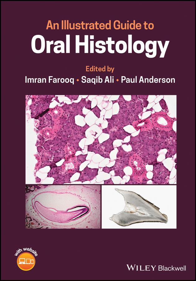
Learn more about the histological presentation of diseased and normal oral tissues with this high definition illustrated dental reference
An Illustrated Guide to Oral Histology delivers a collection of high-definition histological and pathological images, presenting both diseased and normal oral tissues.
The book provides over 200 high-magnification histomicrographs of oral tissues, as well as definitions and explanations of key identifying histological and pathological features of oral tissues. Readers will also benefit from explanations of the clinical significance of particular features, numerous images of ground sections, haemotoxylin- and eosin-stained sections, and electron images. It also includes core topics such as:
Perfect for postgraduate dental students, An Illustrated Guide to Oral Histology will also be useful to undergraduate dental students, and those looking to improve their understanding of the microscopic structure of dental tissues and their pathologies.
Betal enkelt med kort, Klarna, Apple Pay eller Google Pay. Ikke fornøyd? Du har alltid 14 dagers angrerett. Les mer i våre vilkår. Har du spørsmål, send oss en e-post på hello@memmo.org.
Memmo gjør det enklere å studere – uansett hvor du er i verden. Hos oss samler du pensumbøker og smarte studieverktøy på ett og samme sted: sammendrag, quizer, podkaster og flashcards. Og så Ted, din studiekompis som svarer på alt du lurer på. Over 50 000 studenter studerer allerede her – bygget for at du skal lære raskere og stresse mindre.