
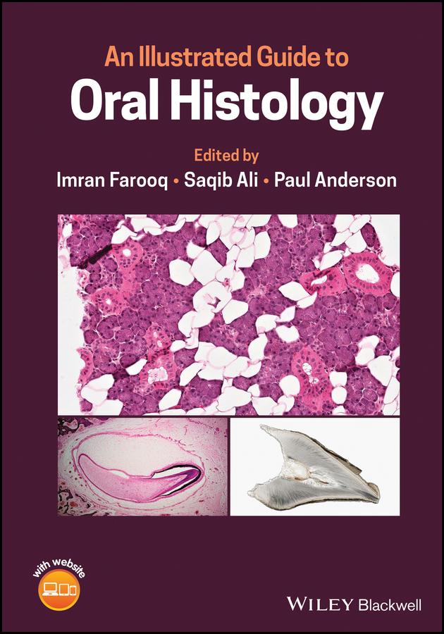
Learn more about the histological presentation of diseased and normal oral tissues with this high definition illustrated dental reference
An Illustrated Guide to Oral Histology delivers a collection of high-definition histological and pathological images, presenting both diseased and normal oral tissues.
The book provides over 200 high-magnification histomicrographs of oral tissues, as well as definitions and explanations of key identifying histological and pathological features of oral tissues. Readers will also benefit from explanations of the clinical significance of particular features, numerous images of ground sections, haemotoxylin- and eosin-stained sections, and electron images. It also includes core topics such as:
Perfect for postgraduate dental students, An Illustrated Guide to Oral Histology will also be useful to undergraduate dental students, and those looking to improve their understanding of the microscopic structure of dental tissues and their pathologies.
Betaal eenvoudig met kaart, Klarna, Apple Pay of Google Pay. Niet tevreden? Je hebt altijd 14 dagen bedenktijd. Lees meer in onze voorwaarden. Heb je vragen, mail ons dan via hello@memmo.org.
Memmo maakt studeren makkelijker – waar je ook bent ter wereld. Wij brengen je cursusboeken en slimme studietools samen op één plek: samenvattingen, quizzen, podcasts en flashcards. En Ted, je studievriend die antwoord geeft op alles wat je je afvraagt. Meer dan 50.000 studenten studeren hier al – gebouwd om je sneller te laten leren en minder stress te geven.