
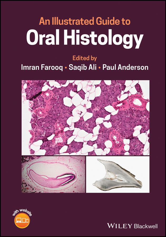
Learn more about the histological presentation of diseased and normal oral tissues with this high definition illustrated dental reference
An Illustrated Guide to Oral Histology delivers a collection of high-definition histological and pathological images, presenting both diseased and normal oral tissues.
The book provides over 200 high-magnification histomicrographs of oral tissues, as well as definitions and explanations of key identifying histological and pathological features of oral tissues. Readers will also benefit from explanations of the clinical significance of particular features, numerous images of ground sections, haemotoxylin- and eosin-stained sections, and electron images. It also includes core topics such as:
Perfect for postgraduate dental students, An Illustrated Guide to Oral Histology will also be useful to undergraduate dental students, and those looking to improve their understanding of the microscopic structure of dental tissues and their pathologies.
Maksa helposti kortilla, Klarnalla, Apple Paylla tai Google Paylla. Etkö ole tyytyväinen? Sinulla on aina 14 päivän palautusoikeus. Lue lisää ehdoistamme. Jos sinulla on kysyttävää, lähetä meille sähköpostia osoitteeseen hello@memmo.org.
Memmo tekee opiskelusta helpompaa – missä päin maailmaa ikinä oletkin. Meillä yhdistät kurssikirjat ja fiksut opiskelutyökalut yhteen paikkaan: tiivistelmät, visat, podcastit ja muistikortit. Ja sitten Ted, opiskelukaverisi, joka vastaa kaikkeen, mitä ikinä mietitkin. Yli 50 000 opiskelijaa opiskelee jo täällä – rakennettu auttamaan sinua oppimaan nopeammin ja stressaamaan vähemmän.