
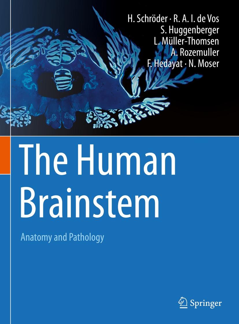
The human brainstem has long been a neglected area in clinical medicine. This is shown by the fact that there is no introductory book on the neuroanatomy and pathology of this region.
This book is intended to introduce the reader to the neuroanatomy of the human brainstem and combines an atlas with detailed information on the individual structures. The atlas features a state-of-the-art magnetic resonance imaging series, histological specimens (Darrow Red and Campbell staining) and a plastinate-based topographical part, which allows direct comparison of histological and topographical findings with neuroimaging. In addition, the reader is guided along the brainstem neuromer model through the human brainstem and learns about the functional properties of the individual structures of the brainstem. Where appropriate, peripheral targets of brainstem structures are illustrated and explained. Furthermore, each chapter covers the most important neurological disorders affecting the brainstem.
This book aims to demonstrate that sound anatomical knowledge is required to understand brainstem pathology. It will particularly help those new to the field to better understand the complex anatomy of the human brainstem and will be useful to basic and clinical neuroscientists alike.
Pay easily by card, Klarna, Apple Pay or Google Pay. Not happy? You always have a 14-day money-back guarantee. Read more in our terms. If you have any questions, email us at hello@memmo.org.
Memmo makes studying easier – wherever you are in the world. We bring your course books and smart study tools together in one place: summaries, quizzes, podcasts and flashcards. Plus Ted, your study buddy who answers anything you wonder. Over 75,000 students already study here – built to help you learn faster and stress less.