
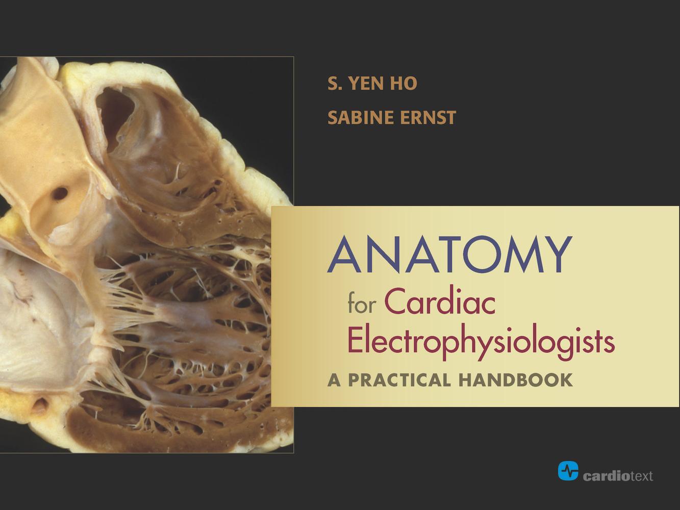
This highly visual handbook integrates cardiac anatomy and the state-of-the-art imaging techniques used in today's catheter or electrophysiology laboratory, guiding readers to a comprehensive understanding of both normal cardiac anatomy and the structures associated with complex heart disease.
Well organized, easily navigable, and superbly illustrated in a landscape format, this unique text invites the reader on a visual intracardiac journey via stunning images and schematic illustrations, including such imaging modalities as computed tomography, magnetic resonance imaging, ultrasound, radiography, and 3D mapping. Each chapter couples the electrophysiology perspective with detailed descriptions of the anatomic features relevant to a wide variety of arrhythmias, including:
Supraventricular tachycardias
Atrial fibrillation
Ventricular arrhythmias
With an overview of general cardiac anatomy, congenital malformations, standard catheter positioning, and potential pitfalls, Anatomy for Cardiac Electrophysiologists provides a solid foundation and quick reference for trainees as they prepare for the realities of the catheter laboratory as well as an excellent refresher for experienced operators.
Pay easily by card, Klarna, Apple Pay or Google Pay. Not happy? You always have a 14-day money-back guarantee. Read more in our terms. If you have any questions, email us at hello@memmo.org.
Memmo makes studying easier – wherever you are in the world. We bring your course books and smart study tools together in one place: summaries, quizzes, podcasts and flashcards. Plus Ted, your study buddy who answers anything you wonder. Over 75,000 students already study here – built to help you learn faster and stress less.
You might also like
Buy the book0