
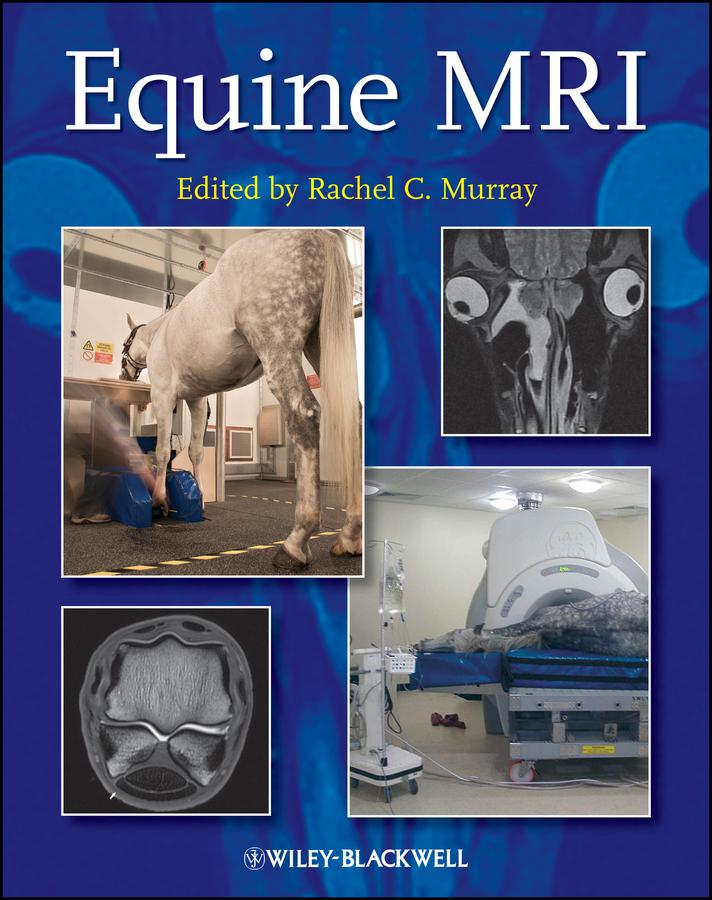
Divided into the following four sections, the book presents key information based on previous validation work and clinical practice:
- Principles of MRI, including the practicalities of image acquisition and interpretation
- Normal MRI anatomy and normal variations
- Different types of pathological change
- Options for clinical management and prognosis for different conditions
MRI is a rapidly expanding area in veterinary medicine that confers detailed, three-dimensional information on both bone and soft tissue. Expanding clinical knowledge, improvements in technology, and practical application of MRI to the standing and recumbent horse means this useful imaging modality has become an integral and essential part of the diagnostic evaluation in lameness and is a realistic option for investigation of ophthalmological, neurological and cranial pathology.
Equine MRI enables readers to understand the best ways to achieve good quality images, and provides a detailed explanation of the problems that may occur.
With close to 950 normal and abnormal images, this book offers considerable detail and examples of both common and uncommon problems, making it a great reference for equine veterinarians, veterinary students, specialists in equine surgery, and specialists in veterinary imaging.
Pay easily by card, Klarna, Apple Pay or Google Pay. Not happy? You always have a 14-day money-back guarantee. Read more in our terms. If you have any questions, email us at hello@memmo.org.
Memmo makes studying easier – wherever you are in the world. We bring your course books and smart study tools together in one place: summaries, quizzes, podcasts and flashcards. Plus Ted, your study buddy who answers anything you wonder. Over 50,000 students already study here – built to help you learn faster and stress less.