
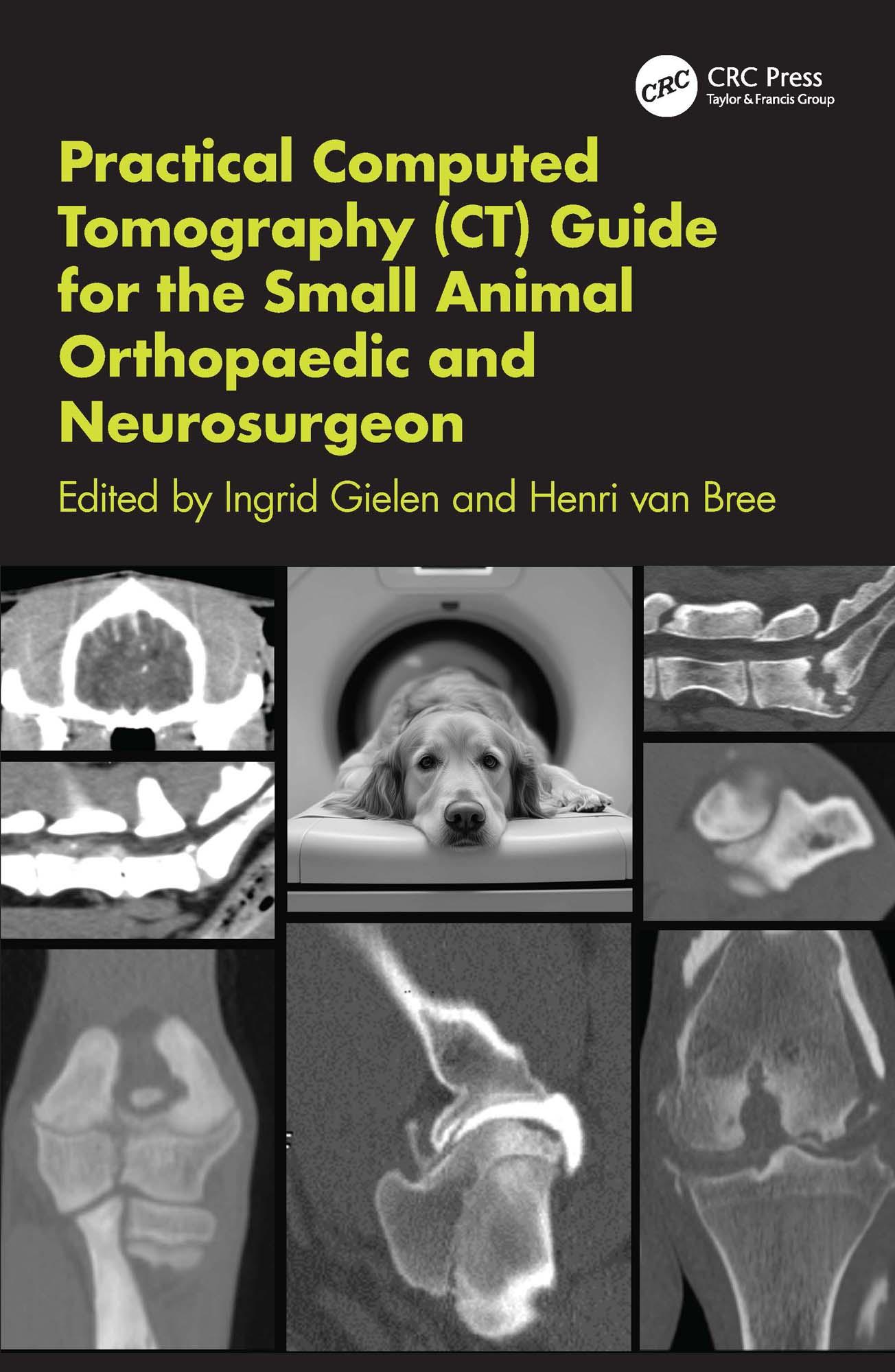
More clinics and large practices are investing in Computed Tomography (CT) equipment, meaning veterinary surgeons are often confronted with unfamiliar technical challenges like positioning and handling of the equipment. Even after obtaining the CT images, the interpretation can often be confusing, particularly when surgeons are not used to reading axial and transverse images.
Split into two parts, looking first at the principles of CT and then drilling down to specific procedures by body region, this book is packed with over 200 high quality images, practical protocols and easy-to-locate information. It provides much needed support in choosing the right equipment, providing the exact protocol and coming to a final diagnosis.
Pay easily by card, Klarna, Apple Pay or Google Pay. Not happy? You always have a 14-day money-back guarantee. Read more in our terms. If you have any questions, email us at hello@memmo.org.
Memmo makes studying easier – wherever you are in the world. We bring your course books and smart study tools together in one place: summaries, quizzes, podcasts and flashcards. Plus Ted, your study buddy who answers anything you wonder. Over 75,000 students already study here – built to help you learn faster and stress less.