
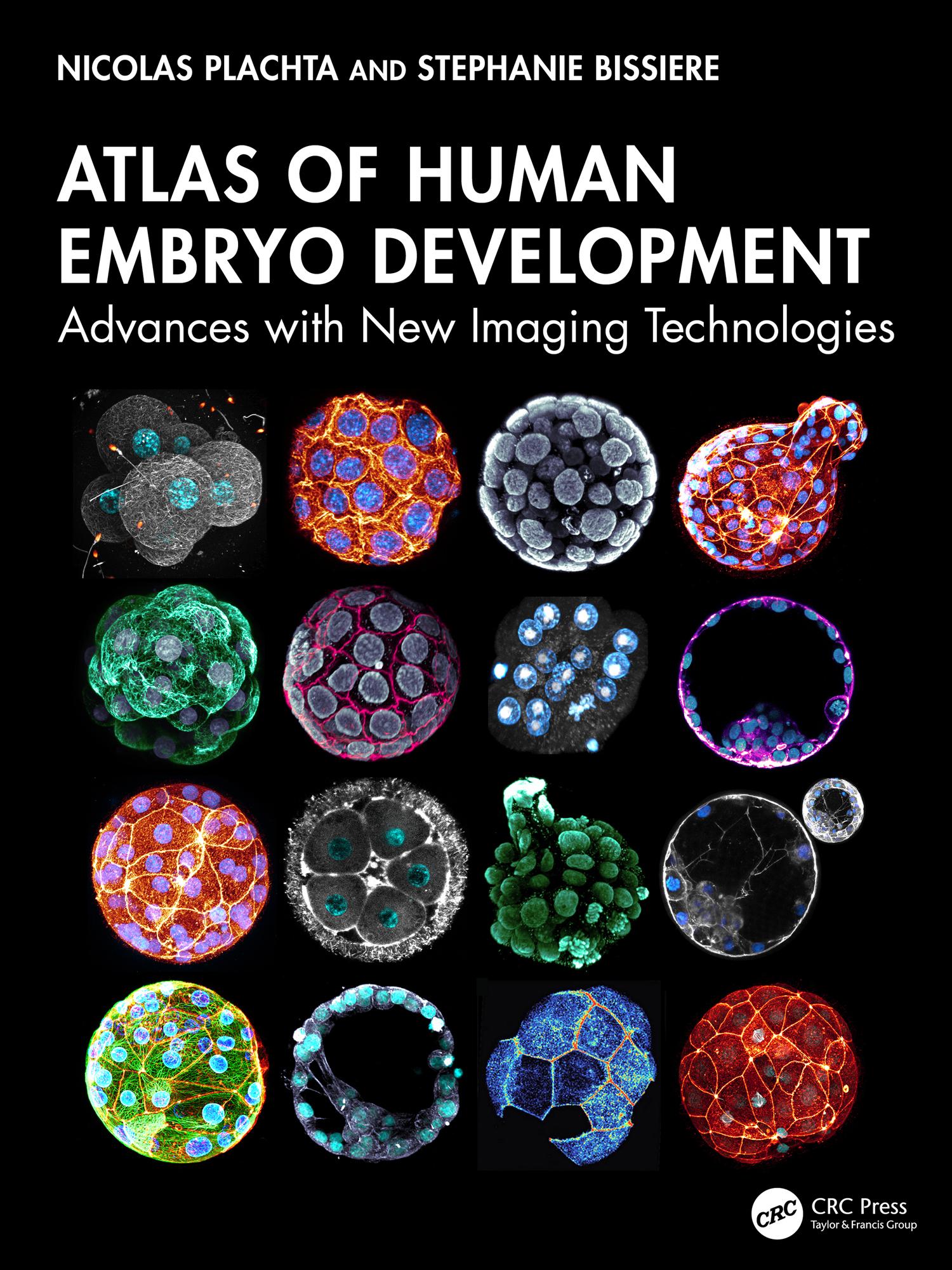
This important and exciting atlas shows with unprecedented resolution the development of the human embryo from fertilization to the end of pre-implantation development. Technology can now capture dynamic changes at the subcellular level that drive embryogenesis and development, allowing the whole team in reproductive medicine to move on from traditional imaging of fixed specimens or classic light microscopy to seeing, as if in 3D and real time, the process of fertilization and development of the human embryo – and of developmental defects. The human embryo research documented here will be of paramount importance for reproductive health, offering the chance to advance fertility treatments, genetic disease prevention, and our understanding of early human development.
Pay easily by card, Klarna, Apple Pay or Google Pay. Not happy? You always have a 14-day money-back guarantee. Read more in our terms. If you have any questions, email us at hello@memmo.org.
Memmo makes studying easier – wherever you are in the world. We bring your course books and smart study tools together in one place: summaries, quizzes, podcasts and flashcards. Plus Ted, your study buddy who answers anything you wonder. Over 75,000 students already study here – built to help you learn faster and stress less.