
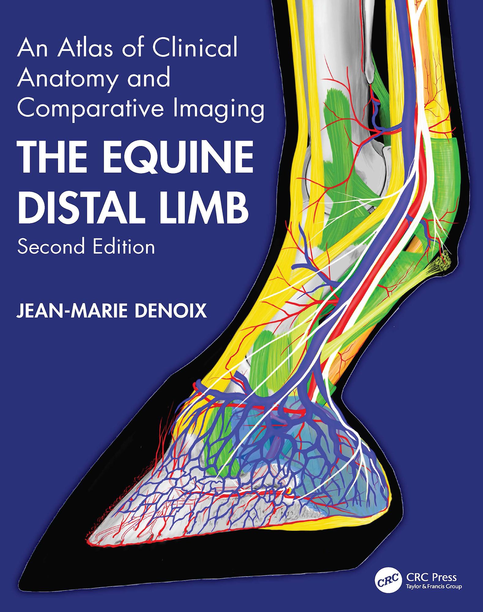
Jean-Marie Denoix is the world’s leading equine musculoskesletal system anatomist and has become one of the foremost equine diagnostic ultrasonographers. There is therefore nobody better to compile a reference atlas of the clinical anatomy of the foot, pastern and fetlock, correlated with images obtained by radiography, diagnostic ultrasonography and magnetic resonance imaging. Advanced imaging techniques require in-depth knowledge of anatomy for accurate interpretation and, especially when using magnetic resonance imaging, this must be a three-dimensional concept of anatomy.
This new edition replaces ultrasound images and most of the radiographic and MRI images with new, updated versions and adds brand new images of extraordinarily high quality. The multiple views of each area of the distal limb provide an extremely detailed evaluation, while every part opens with an anatomical drawing by the author. Each double-page spread deals with a single dissection viewed by means of colour photographs, labelled B&W equivalents, plus x-rays, ultrasound and MRI scans as required.
Diagnosis and management of distal limb lameness require a precise knowledge of the functional anatomy and biomechanics of the equine distal joints, ligaments and tendons, presented in the last chapter. The atlas is designed for maximum clarity using a generous page size and is essential for anybody involved in detailed anatomical study, complex lameness evaluation or advanced imaging techniques.
Pay easily by card, Klarna, Apple Pay or Google Pay. Not happy? You always have a 14-day money-back guarantee. Read more in our terms. If you have any questions, email us at hello@memmo.org.
Memmo makes studying easier – wherever you are in the world. We bring your course books and smart study tools together in one place: summaries, quizzes, podcasts and flashcards. Plus Ted, your study buddy who answers anything you wonder. Over 75,000 students already study here – built to help you learn faster and stress less.