
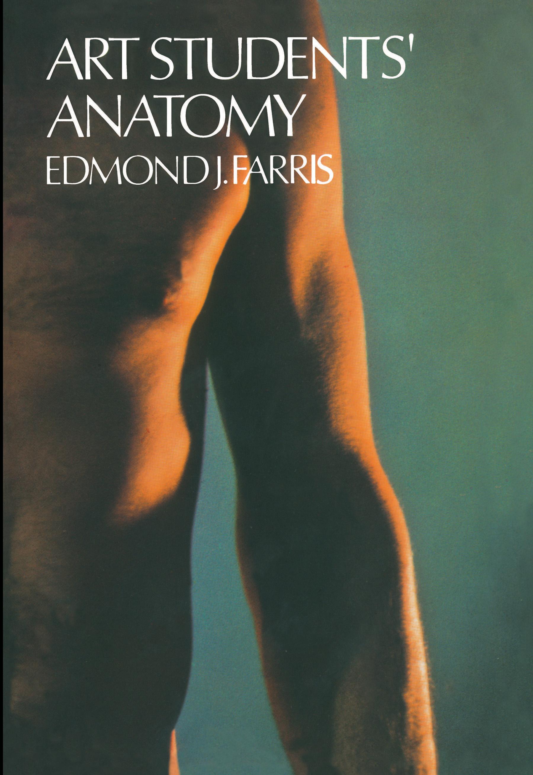
For art students working with the human figure, this comprehensive study of the bones, muscles, and surface forms of the living body will be one of the most useful (and most used) additions they can make to their private libraries. More than 150 illustrations, mostly full-page photographs and labeled sketches of undraped male and female bodies, provide the reader with anatomical studies of unrivaled clarity and unquestioned accuracy.
After an introduction covering the proportions of the adult male, the adult female, and the infant at various ages, the author devotes 50 pages to the human skeletal system. Besides pictures and detailed drawings of the major bones of the body, he also includes x-rays showing the bone structure of the hand and foot and the movements of the shoulder, elbow, and knee joints.
A section on the muscular system follows, including reproductions of the remarkable Albinus engravings ("The most beautiful and among the most accurate anatomical figures ever published." — Charles Singer), 36 photographs and labeled sketches of living models, and seven drawings showing the attachments of muscles to the skeleton. The book concludes with a number of poses and action photographs illustrating surface anatomy in various actions such as dancing and throwing a ball.
This is one of the few and perhaps the best of those books that teach anatomy using chiefly living objects for their illustrative work. By doing so, it fills an urgent need, for most art students cannot afford living models or expensive courses in anatomy. Now, however, they can use this practical and inexpensive home-study course to achieve a clearer insight into the complicated mechanism of the human body, as simplified by Dr. Farris.
After an introduction covering the proportions of the adult male, the adult female, and the infant at various ages, the author devotes 50 pages to the human skeletal system. Besides pictures and detailed drawings of the major bones of the body, he also includes x-rays showing the bone structure of the hand and foot and the movements of the shoulder, elbow, and knee joints.
A section on the muscular system follows, including reproductions of the remarkable Albinus engravings ("The most beautiful and among the most accurate anatomical figures ever published." — Charles Singer), 36 photographs and labeled sketches of living models, and seven drawings showing the attachments of muscles to the skeleton. The book concludes with a number of poses and action photographs illustrating surface anatomy in various actions such as dancing and throwing a ball.
This is one of the few and perhaps the best of those books that teach anatomy using chiefly living objects for their illustrative work. By doing so, it fills an urgent need, for most art students cannot afford living models or expensive courses in anatomy. Now, however, they can use this practical and inexpensive home-study course to achieve a clearer insight into the complicated mechanism of the human body, as simplified by Dr. Farris.
Pages: 159
Category: Konst, Design & Fotografi
e-ISBN: 9780486158105
Shipping weight: 1.2 kg
Ships in: 1-2 days
Pay easily by card, Klarna, Apple Pay or Google Pay. Not happy? You always have a 14-day money-back guarantee. Read more in our terms. If you have any questions, email us at hello@memmo.org.
Memmo makes studying easier – wherever you are in the world. We bring your course books and smart study tools together in one place: summaries, quizzes, podcasts and flashcards. Plus Ted, your study buddy who answers anything you wonder. Over 50,000 students already study here – built to help you learn faster and stress less.
You might also like
Buy the book0