
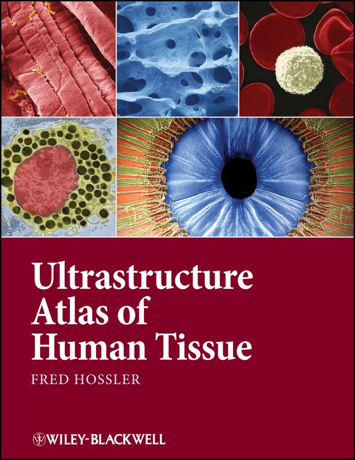
Ultrastructure Atlas of Human Tissues presents a variety of scanning and transmission electron microscope images of the major systems of the human body. Photography with the electron microscope records views of the intricate substructures and microdesigns of objects and tissues, and reveals details within them inaccessible to the naked eye or light microscope. Many of these views have significance in understanding normal structure and function, as well as disease processes. This book offers a unique and comprehensive look at the structure and function of tissues at the subcellular and molecular level, an important perspective in understanding and combating diseases.
• Presents the major systems of the human body through scanning and transmission electron microscope images
• Has images prepared almost exclusively from human tissues
• Includes electron micrographs of common pathologies such as fibrotic and emphysemic lung, kidney stones, sickle cell anemia, and skin parasites
• Contains sets of 3D images in most chapters
Betala smidigt med kort, Klarna, Apple Pay eller Google Pay. Är du inte nöjd har du alltid 14 dagars ångerrätt. Läs mer i våra villkor. Har du några frågor, mejla oss på hello@memmo.org.
Memmo gör det enklare att plugga – var du än är i världen. Hos oss samlar du kursböcker och smarta studieverktyg på ett och samma ställe: sammanfattningar, quiz, poddar och flashcards. Och så Ted, din studiekompis som svarar på allt du undrar. Över 50 000 studenter pluggar redan här – byggt för att du ska lära dig snabbare och stressa mindre.