
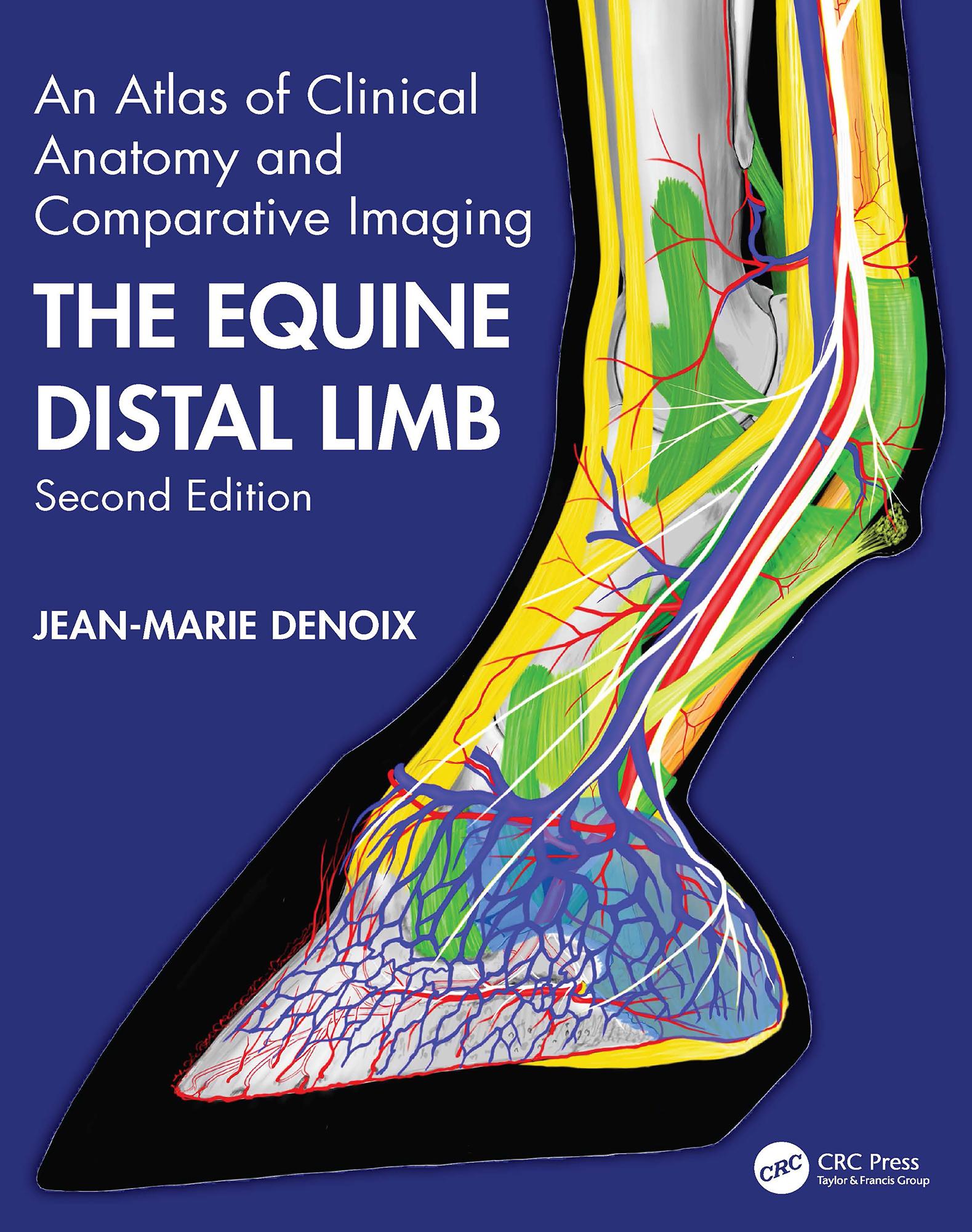
Jean-Marie Denoix is the world’s leading equine musculoskesletal system anatomist and has become one of the foremost equine diagnostic ultrasonographers. There is therefore nobody better to compile a reference atlas of the clinical anatomy of the foot, pastern and fetlock, correlated with images obtained by radiography, diagnostic ultrasonography and magnetic resonance imaging. Advanced imaging techniques require in-depth knowledge of anatomy for accurate interpretation and, especially when using magnetic resonance imaging, this must be a three-dimensional concept of anatomy.
This new edition replaces ultrasound images and most of the radiographic and MRI images with new, updated versions and adds brand new images of extraordinarily high quality. The multiple views of each area of the distal limb provide an extremely detailed evaluation, while every part opens with an anatomical drawing by the author. Each double-page spread deals with a single dissection viewed by means of colour photographs, labelled B&W equivalents, plus x-rays, ultrasound and MRI scans as required.
Diagnosis and management of distal limb lameness require a precise knowledge of the functional anatomy and biomechanics of the equine distal joints, ligaments and tendons, presented in the last chapter. The atlas is designed for maximum clarity using a generous page size and is essential for anybody involved in detailed anatomical study, complex lameness evaluation or advanced imaging techniques.
Płać wygodnie kartą, Klarną, Apple Pay lub Google Pay. Nie jesteś zadowolony? Zawsze masz 14-dniową gwarancję zwrotu pieniędzy. Więcej przeczytasz w naszych warunkach. Masz pytania? Napisz do nas na hello@memmo.org.
Memmo ułatwia naukę – gdziekolwiek jesteś na świecie. U nas znajdziesz podręczniki i sprytne narzędzia do nauki w jednym miejscu: streszczenia, quizy, podcasty i fiszki. A do tego Ted, Twój kumpel do nauki, który odpowie na wszystko, co Cię nurtuje. Ponad 50 000 studentów już tu się uczy – stworzone, byś uczył się szybciej i mniej stresował.