
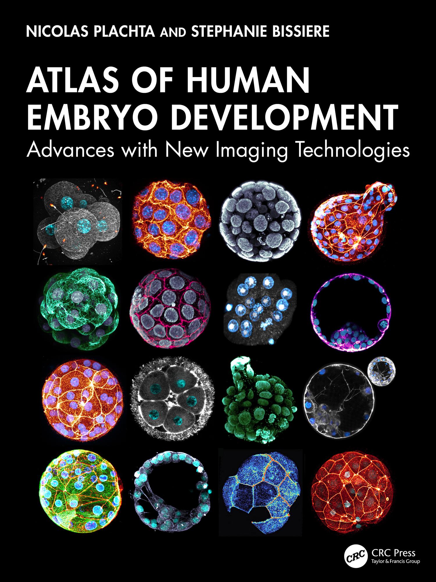
This important and exciting atlas shows with unprecedented resolution the development of the human embryo from fertilization to the end of pre-implantation development. Technology can now capture dynamic changes at the subcellular level that drive embryogenesis and development, allowing the whole team in reproductive medicine to move on from traditional imaging of fixed specimens or classic light microscopy to seeing, as if in 3D and real time, the process of fertilization and development of the human embryo – and of developmental defects. The human embryo research documented here will be of paramount importance for reproductive health, offering the chance to advance fertility treatments, genetic disease prevention, and our understanding of early human development.
Betal enkelt med kort, Klarna, Apple Pay eller Google Pay. Ikke fornøyd? Du har alltid 14 dagers angrerett. Les mer i våre vilkår. Har du spørsmål, send oss en e-post på hello@memmo.org.
Memmo gjør det enklere å studere – uansett hvor du er i verden. Hos oss samler du pensumbøker og smarte studieverktøy på ett og samme sted: sammendrag, quizer, podkaster og flashcards. Og så Ted, din studiekompis som svarer på alt du lurer på. Over 50 000 studenter studerer allerede her – bygget for at du skal lære raskere og stresse mindre.