
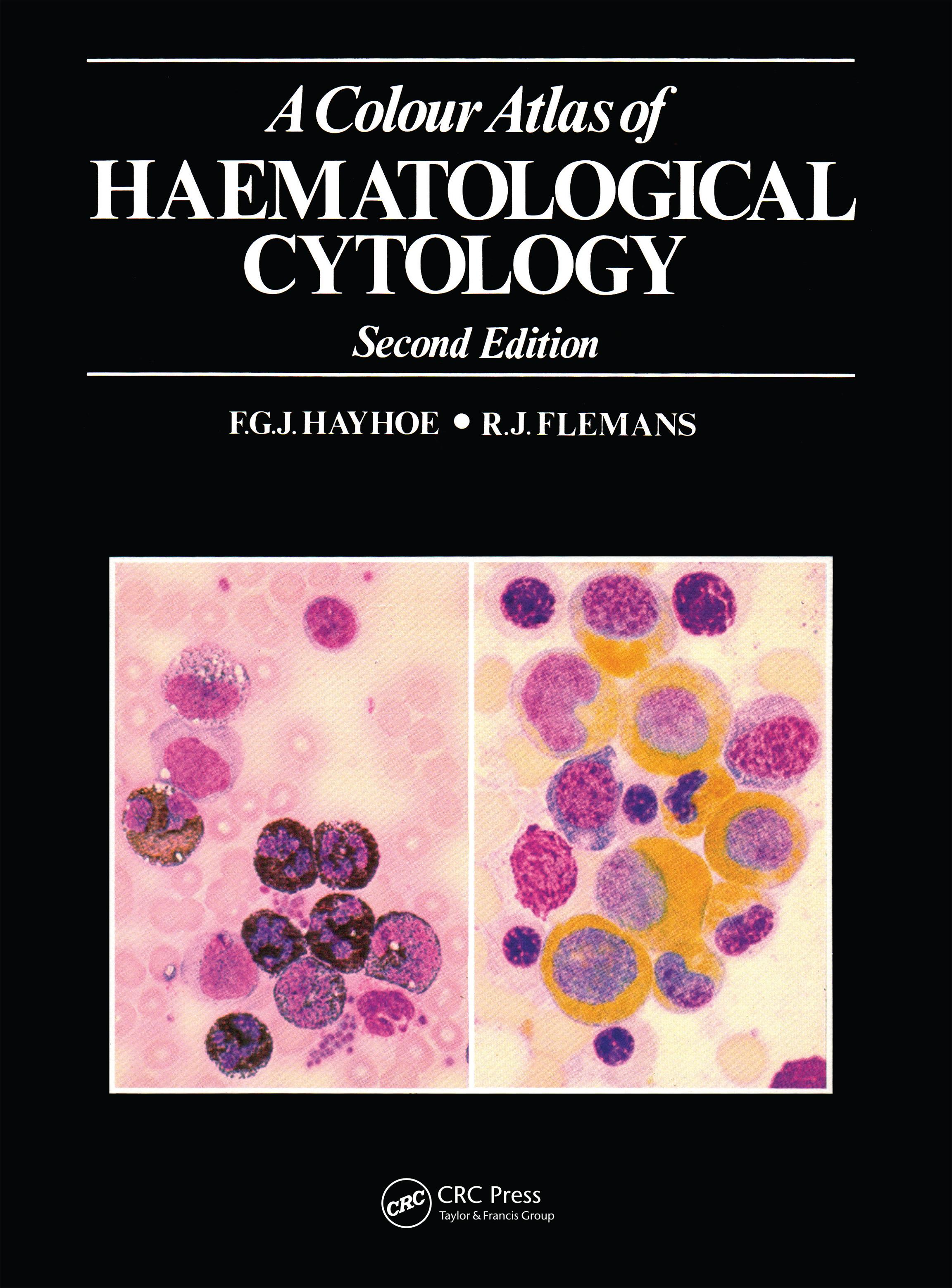
The second edition of this atlas has been revised and greatly en-larged to take account of recent diagnostic advances in the field of haematology. It contains 750 color photomicrographs of common and less common cell types, and is intended to supplement the per-sonal study of preparations under the microscope.
The photographs are grouped into five sections covering red cells and their precursors; nonerythroid cells of myeloid origin; lympho-cytes, plasma cells and their derivatives and precursors; miscella-neous cells of blood and marrow, including foreign cells and para-sites; and the imprint cytology of lymph nodes and spleen. Each section is preceded by a brief introduction, and an appendix pro-vides technical details of staining methods. The text accompanying the illustrations serves chiefly to identify the cells, but also gives explanations of certain unusual appearances and the cytochemical reactions where necessary.
A Colour Atlas of Haematological Cytology will be useful to students of medicine and medical laboratory science, as well as to doctors and technicians specializing in the diagnosis of haematological disease.
Betal enkelt med kort, Klarna, Apple Pay eller Google Pay. Ikke fornøyd? Du har alltid 14 dagers angrerett. Les mer i våre vilkår. Har du spørsmål, send oss en e-post på hello@memmo.org.
Memmo gjør det enklere å studere – uansett hvor du er i verden. Hos oss samler du pensumbøker og smarte studieverktøy på ett og samme sted: sammendrag, quizer, podkaster og flashcards. Og så Ted, din studiekompis som svarer på alt du lurer på. Over 50 000 studenter studerer allerede her – bygget for at du skal lære raskere og stresse mindre.
Du vil kanskje også like
Kjøp boken0