
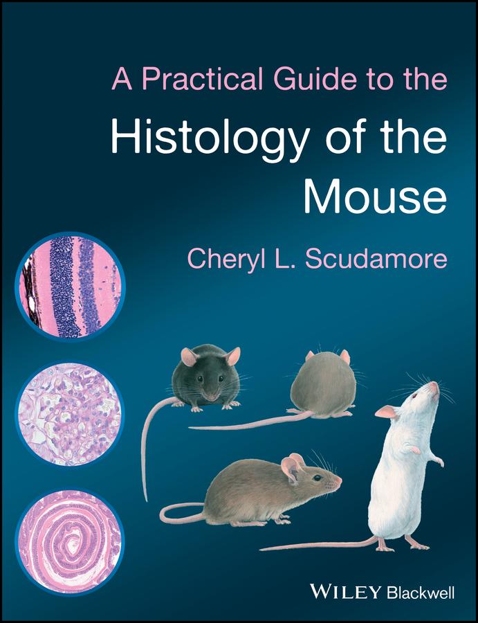
This book is aimed at veterinary and medical pathologists who are unfamiliar with mouse tissues and scientists who wish to evaluate their own mouse models. It provides practical guidance on the collection, sampling and analysis of mouse tissue samples in order to maximize the information that can be gained from these tissues. As well as illustrating the normal microscopic anatomy of the mouse, the book also describes and explains the common anatomic variations, artefacts associated with tissue collection and background lesions to help the scientist to distinguish these changes from experimentally- induced lesions.
This will be an essential bench-side companion for researchers and practitioners looking for an accessible and well-illustrated guide to mouse pathology.
- Written by experienced pathologists and specifically tailored to the needs of scientists and histologists
- Full colour throughout
- Provides advice on sampling tissues, necropsy and recording data
- Includes common anatomic variations, background lesions and artefacts which will help non-experts understand whether histologic variations seen are part of the normal background or related to their experimental manipulation
Betaal eenvoudig met kaart, Klarna, Apple Pay of Google Pay. Niet tevreden? Je hebt altijd 14 dagen bedenktijd. Lees meer in onze voorwaarden. Heb je vragen, mail ons dan via hello@memmo.org.
Memmo maakt studeren makkelijker – waar je ook bent ter wereld. Wij brengen je cursusboeken en slimme studietools samen op één plek: samenvattingen, quizzen, podcasts en flashcards. En Ted, je studievriend die antwoord geeft op alles wat je je afvraagt. Meer dan 50.000 studenten studeren hier al – gebouwd om je sneller te laten leren en minder stress te geven.