
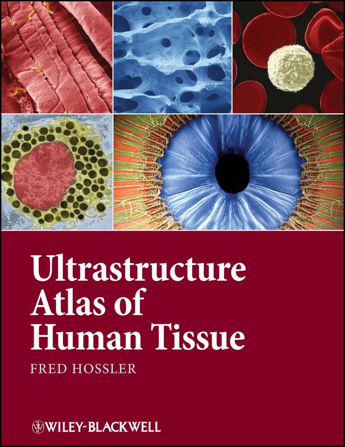
Ultrastructure Atlas of Human Tissues presents a variety of scanning and transmission electron microscope images of the major systems of the human body. Photography with the electron microscope records views of the intricate substructures and microdesigns of objects and tissues, and reveals details within them inaccessible to the naked eye or light microscope. Many of these views have significance in understanding normal structure and function, as well as disease processes. This book offers a unique and comprehensive look at the structure and function of tissues at the subcellular and molecular level, an important perspective in understanding and combating diseases.
• Presents the major systems of the human body through scanning and transmission electron microscope images
• Has images prepared almost exclusively from human tissues
• Includes electron micrographs of common pathologies such as fibrotic and emphysemic lung, kidney stones, sickle cell anemia, and skin parasites
• Contains sets of 3D images in most chapters
Betaal eenvoudig met kaart, Klarna, Apple Pay of Google Pay. Niet tevreden? Je hebt altijd 14 dagen bedenktijd. Lees meer in onze voorwaarden. Heb je vragen, mail ons dan via hello@memmo.org.
Memmo maakt studeren makkelijker – waar je ook bent ter wereld. Wij brengen je cursusboeken en slimme studietools samen op één plek: samenvattingen, quizzen, podcasts en flashcards. En Ted, je studievriend die antwoord geeft op alles wat je je afvraagt. Meer dan 50.000 studenten studeren hier al – gebouwd om je sneller te laten leren en minder stress te geven.