
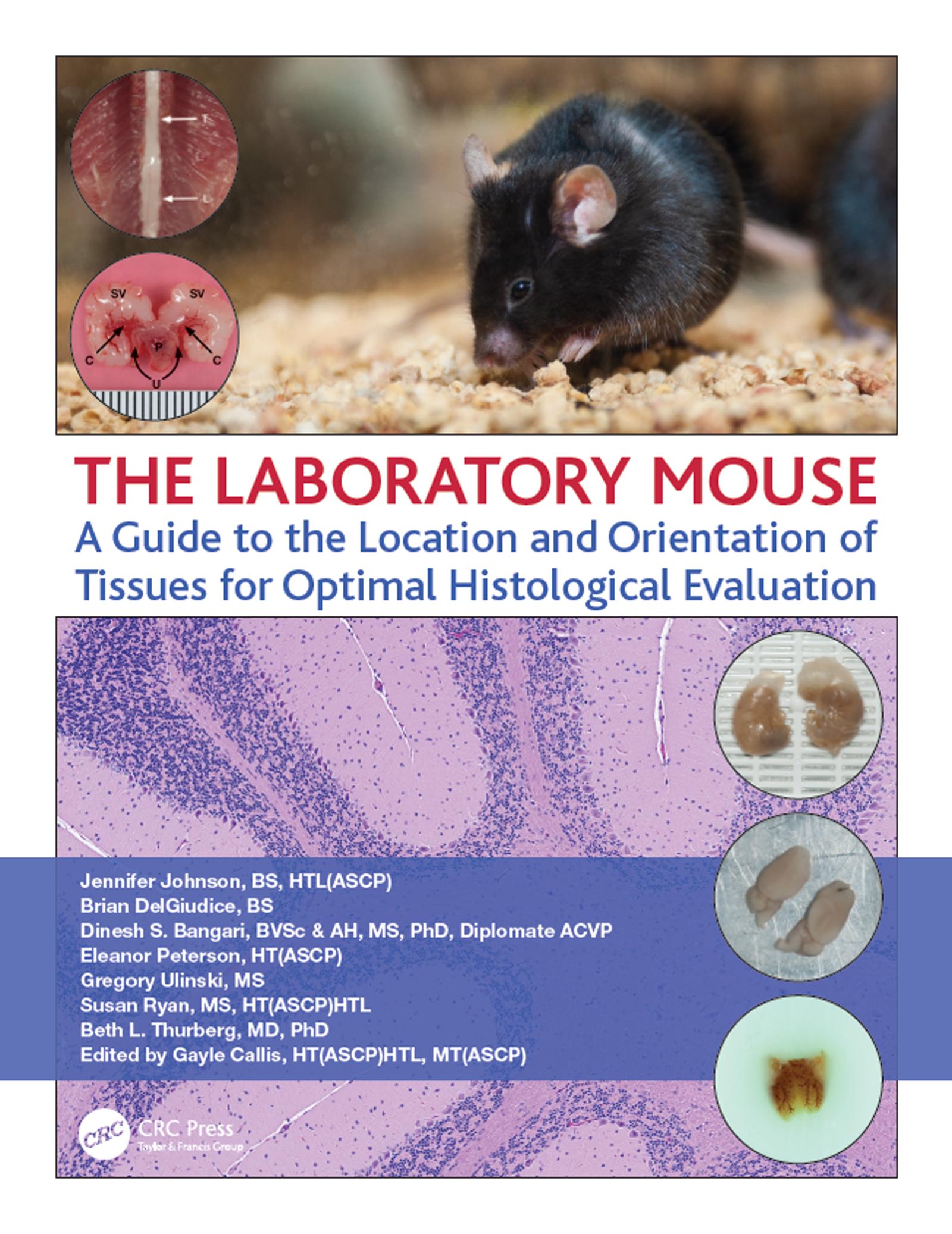
Key features:
- High quality full color photographs and descriptive texts on the location and removal of the organs from the mouse
- Instructive methods and clear visuals for trimming and orienting the organs for paraffin histology to obtain the best possible sections for analysis
- Full color photomicrographs of the resulting section for each organ stained with hematoxylin and eosin demonstrating important features and landmarks for the histologist to ensure the optimal area for analysis is achieved
- All in one, easy to use guide organized by individual organs of the laboratory mouse
-
Spiralbound for easy reference in the lab
This "one-stop" guide offers an essential resource for any academic, research or development operation where mouse necropsy and/or histology are performed. Connecting the reader ‘from the mouse to the microscope’, it provides a detailed guide for locating, trimming, orientating and embedding of the most frequently investigated tissues collected in the laboratory mouse. It shows where the organs reside in the mouse, how to trim and embed them as well as the resulting optimal sections. This guide brings together the wealth of scattered information into one high-quality text, the emphasis is on providing knowledge that will help histologists and scientists get better results in any downstream assays where ideal sections are needed.
Betaal eenvoudig met kaart, Klarna, Apple Pay of Google Pay. Niet tevreden? Je hebt altijd 14 dagen bedenktijd. Lees meer in onze voorwaarden. Heb je vragen, mail ons dan via hello@memmo.org.
Memmo maakt studeren makkelijker – waar je ook bent ter wereld. Wij brengen je cursusboeken en slimme studietools samen op één plek: samenvattingen, quizzen, podcasts en flashcards. En Ted, je studievriend die antwoord geeft op alles wat je je afvraagt. Meer dan 50.000 studenten studeren hier al – gebouwd om je sneller te laten leren en minder stress te geven.