
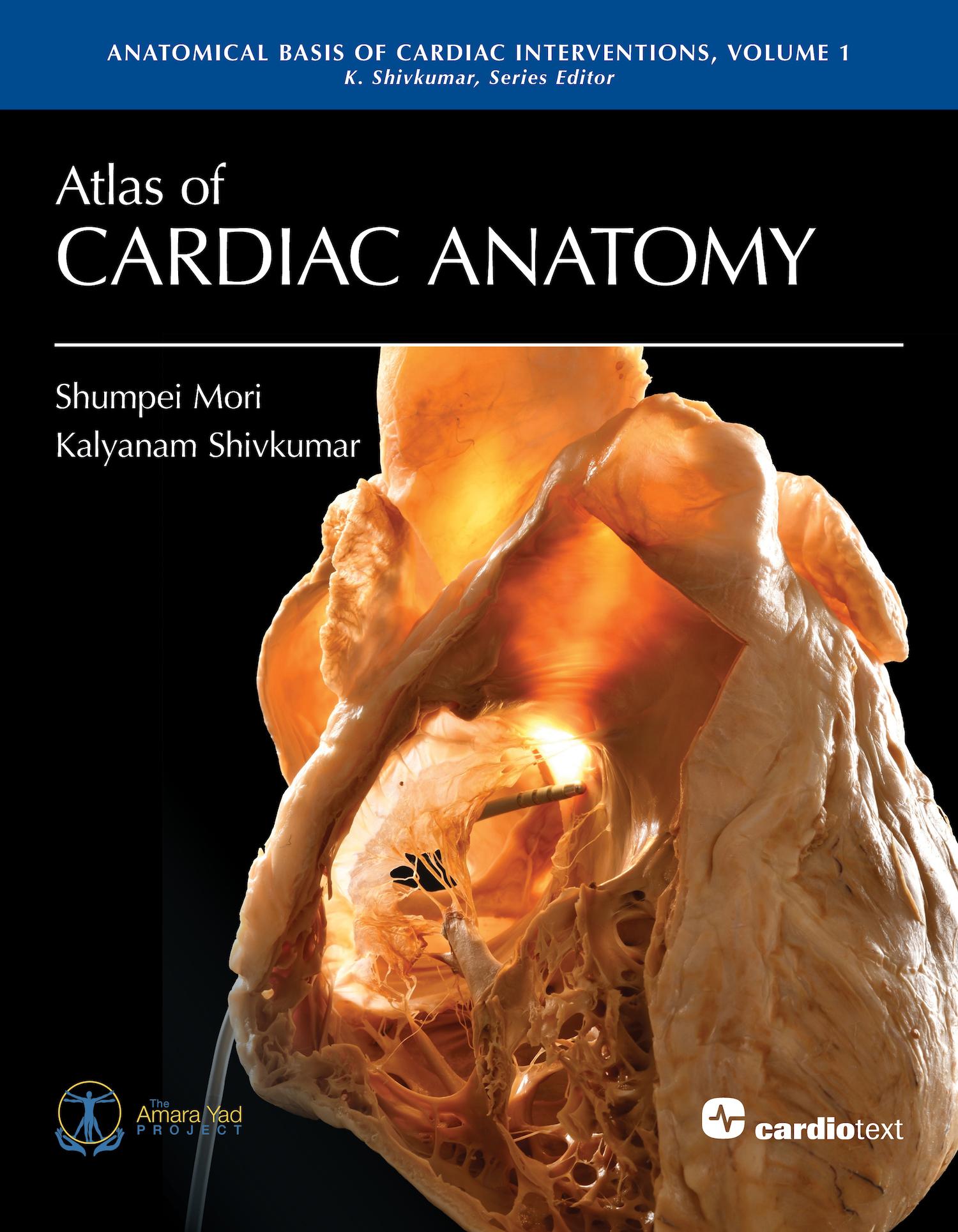
Atlas of Cardiac Anatomy represents a new standard for the study of the human heart.
Drs. Shumpei Mori and Kalyanam Shivkumar have created a visually stunning presentation of more than 200 full-color photographs of the human heart and its adjacent structures.
Atlas of Cardiac Anatomy includes unpublished and restored iconic photographs from the Wallace A. McAlpine Collection at UCLA and new images from the UCLA Cardiac Arrhythmia Center based on the methods and photographic techniques for the study of cardiac anatomy pioneered by Dr. McAlpine.
This first volume serves as a core foundational study of cardiac anatomy and provides a basis for planned atlases edited by Dr. Shivkumar in the series, Anatomical Basis of Cardiac Interventions. Electrophysiology, structural heart disease, imaging, and cardiac surgery are the focus of future atlases.
The authors’ goals are to offer an exceptionally comprehensive resource on cardiac anatomy. It aims to inspire new solutions and provide additional perspectives for skilled interventional and surgical operators. More broadly, it is likely to be useful for anyone caring for patients with cardiac disease.
Includes access to 25 anaglyphs (3D images). 3D glasses required for viewing and are not included.The authors are at the UCLA Cardiac Arrhythmia Center, Los Angeles, California.
Foreword by Francis E. Marchlinski, MD, and William G. Stevenson, MD.
Payez facilement par carte, Klarna, Apple Pay ou Google Pay. Pas satisfait ? Vous avez toujours une garantie de remboursement de 14 jours. En savoir plus dans nos conditions. Si vous avez des questions, envoyez-nous un e-mail à hello@memmo.org.
Memmo facilite tes études, où que tu sois dans le monde. On rassemble tes manuels de cours et des outils d'étude intelligents au même endroit : résumés, quiz, podcasts et flashcards. Et il y a Ted, ton compagnon d'étude qui répond à toutes tes questions. Plus de 50 000 étudiants étudient déjà ici – conçu pour t'aider à apprendre plus vite et à moins stresser.