
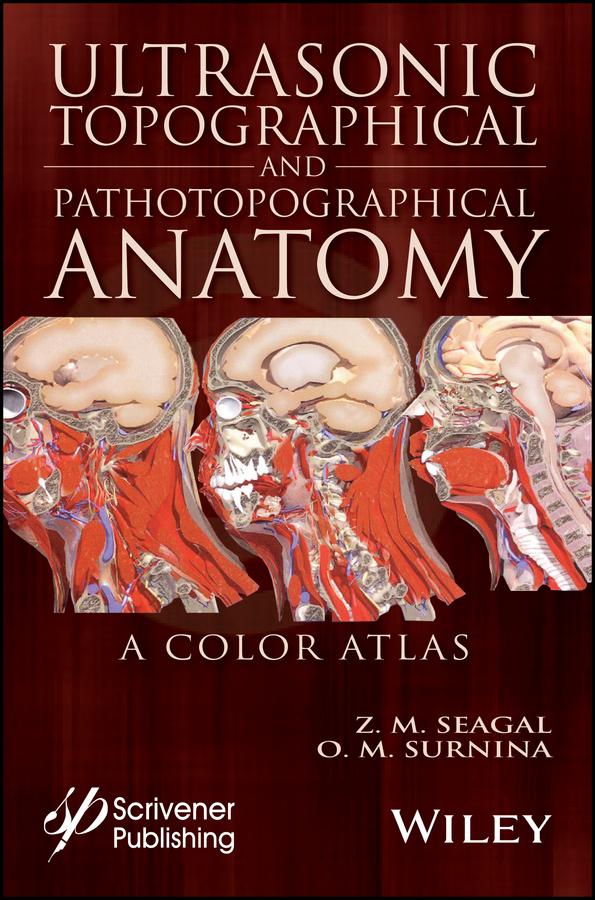
Written by experienced and well-respected physicians and professors, this new all-color volume presents the ultrasonic topographical and pathotopographical anatomy of the body, including the head, neck, chest, anterolateral abdominal wall, abdominal organs, retroperitoneal space, male and female pelvises, and lower extremities.
Specific and non-specific ultrasonic symptoms are suggested for normal and abnormal developmental variants, diffuse and local pathotopographical anatomy. This color atlas contains comparative topographical and pathotopographical data and is the first manual of its kind for students and medical specialists in different areas, including those specializing in medical sonography. The original technology was tested at clinics in patients subjected to ultrasonic monitoring. Because of early detection there were no false-positive or false-negative results. The therapy was effective, and, in some cases, the use of the original method of "seagalography" (optometry and pulsemotorgraphy) has made it possible to develop new methods of treatment and/or to determine the optimal doses of drugs, as well as to develop effective drug complexes for treatment of a given pathology.
This important new volume will be valuable to physicians, junior physicians, medical residents, lecturers in medicine, and medical students alike, either as a textbook or as a reference. It is a must-have for any physician's library.
Payez facilement par carte, Klarna, Apple Pay ou Google Pay. Pas satisfait ? Vous avez toujours une garantie de remboursement de 14 jours. En savoir plus dans nos conditions. Si vous avez des questions, envoyez-nous un e-mail à hello@memmo.org.
Memmo facilite tes études, où que tu sois dans le monde. On rassemble tes manuels de cours et des outils d'étude intelligents au même endroit : résumés, quiz, podcasts et flashcards. Et il y a Ted, ton compagnon d'étude qui répond à toutes tes questions. Plus de 50 000 étudiants étudient déjà ici – conçu pour t'aider à apprendre plus vite et à moins stresser.