
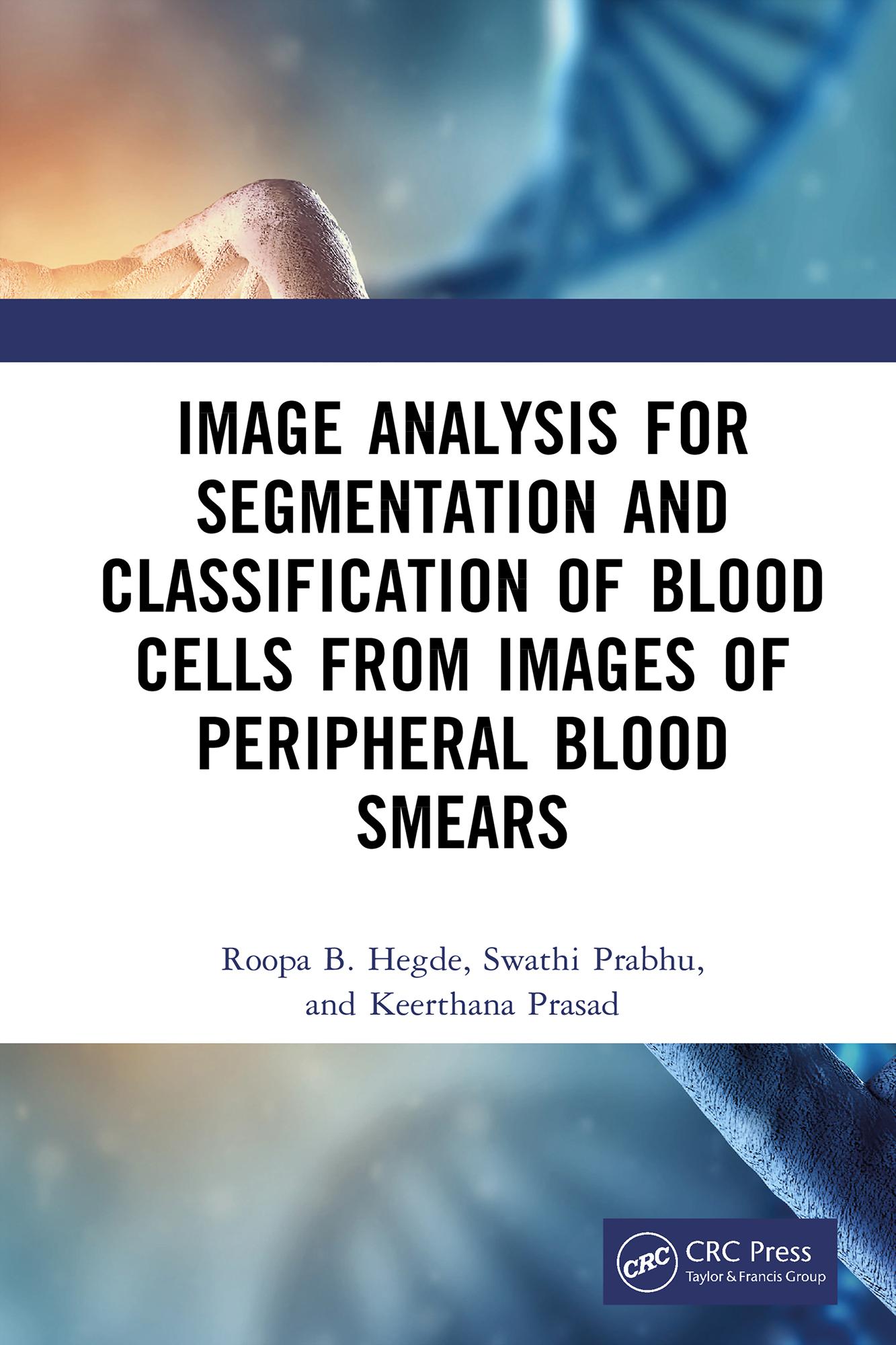
Peripheral blood smear (PBS) analysis is a critical diagnostic tool for diseases including malaria, leukemia, and cancer. This monograph addresses the key challenges associated with image analysis of PBS images and highlights innovative systems that automate the identification of specific pathologies.
It reviews state-of-the-art algorithms for segmentation, color representation and normalization, image enhancement and restoration, and volume visualization, as well as relevant AI and machine learning techniques. The book also explains automated systems and workflows developed to address specific disease classes, supported by detailed case studies.
Key Features:
• Covers fundamental and advanced aspects of image analysis in the hematological field
• Focuses on blood cell segmentation and classification, along with practical guidelines for implementation
• Explores current challenges in integrating these technologies into real-world clinical settings
• Discusses ethical considerations and challenges associated with automation in medical diagnosis
• Includes relevant MATLAB and Python code, along with case studies on the segmentation of erythrocytes (RBCs) and leukocytes (WBCs)
This book is aimed at graduate students and researchers in bioengineering and image processing, as well as clinicians.
Maksa helposti kortilla, Klarnalla, Apple Paylla tai Google Paylla. Etkö ole tyytyväinen? Sinulla on aina 14 päivän palautusoikeus. Lue lisää ehdoistamme. Jos sinulla on kysyttävää, lähetä meille sähköpostia osoitteeseen hello@memmo.org.
Memmo tekee opiskelusta helpompaa – missä päin maailmaa ikinä oletkin. Meillä yhdistät kurssikirjat ja fiksut opiskelutyökalut yhteen paikkaan: tiivistelmät, visat, podcastit ja muistikortit. Ja sitten Ted, opiskelukaverisi, joka vastaa kaikkeen, mitä ikinä mietitkin. Yli 50 000 opiskelijaa opiskelee jo täällä – rakennettu auttamaan sinua oppimaan nopeammin ja stressaamaan vähemmän.