
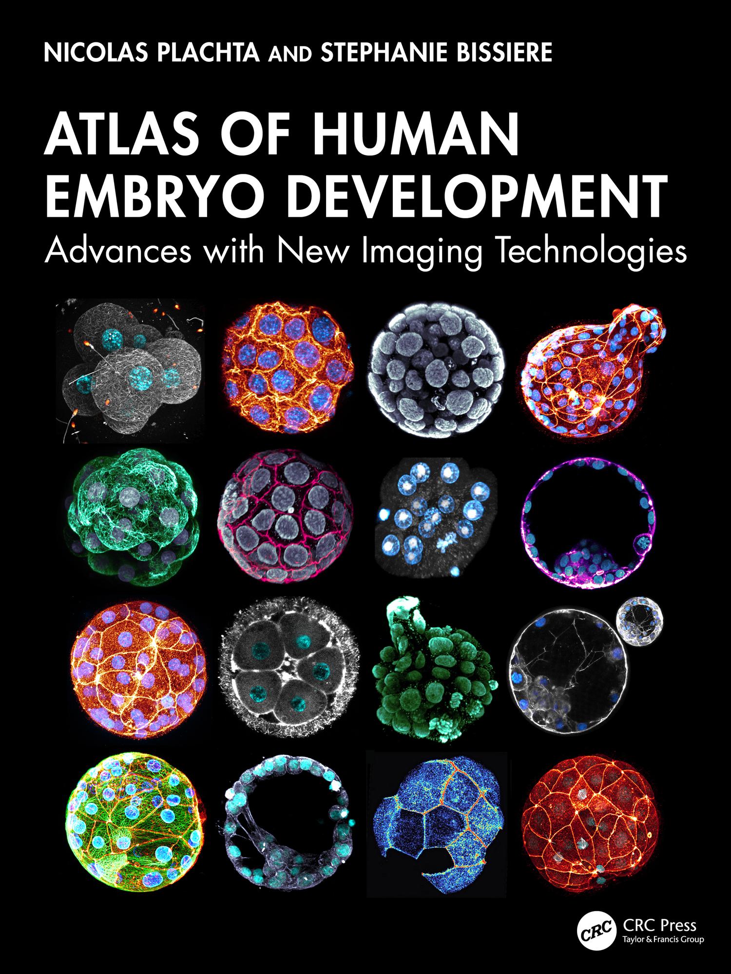
This important and exciting atlas shows with unprecedented resolution the development of the human embryo from fertilization to the end of pre-implantation development. Technology can now capture dynamic changes at the subcellular level that drive embryogenesis and development, allowing the whole team in reproductive medicine to move on from traditional imaging of fixed specimens or classic light microscopy to seeing, as if in 3D and real time, the process of fertilization and development of the human embryo – and of developmental defects. The human embryo research documented here will be of paramount importance for reproductive health, offering the chance to advance fertility treatments, genetic disease prevention, and our understanding of early human development.
Maksa helposti kortilla, Klarnalla, Apple Paylla tai Google Paylla. Etkö ole tyytyväinen? Sinulla on aina 14 päivän palautusoikeus. Lue lisää ehdoistamme. Jos sinulla on kysyttävää, lähetä meille sähköpostia osoitteeseen hello@memmo.org.
Memmo tekee opiskelusta helpompaa – missä päin maailmaa ikinä oletkin. Meillä yhdistät kurssikirjat ja fiksut opiskelutyökalut yhteen paikkaan: tiivistelmät, visat, podcastit ja muistikortit. Ja sitten Ted, opiskelukaverisi, joka vastaa kaikkeen, mitä ikinä mietitkin. Yli 50 000 opiskelijaa opiskelee jo täällä – rakennettu auttamaan sinua oppimaan nopeammin ja stressaamaan vähemmän.