
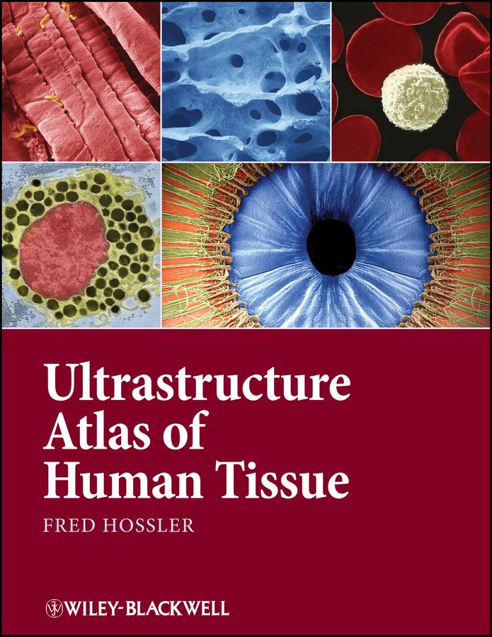
Ultrastructure Atlas of Human Tissues presents a variety of scanning and transmission electron microscope images of the major systems of the human body. Photography with the electron microscope records views of the intricate substructures and microdesigns of objects and tissues, and reveals details within them inaccessible to the naked eye or light microscope. Many of these views have significance in understanding normal structure and function, as well as disease processes. This book offers a unique and comprehensive look at the structure and function of tissues at the subcellular and molecular level, an important perspective in understanding and combating diseases.
• Presents the major systems of the human body through scanning and transmission electron microscope images
• Has images prepared almost exclusively from human tissues
• Includes electron micrographs of common pathologies such as fibrotic and emphysemic lung, kidney stones, sickle cell anemia, and skin parasites
• Contains sets of 3D images in most chapters
Paga facilmente con carta, Klarna, Apple Pay o Google Pay. Non sei soddisfatto? Hai sempre 14 giorni per il rimborso. Leggi di più nei nostri termini. Per qualsiasi domanda, scrivici a hello@memmo.org.
Memmo rende lo studio più facile, ovunque tu sia nel mondo. Qui trovi i tuoi libri di testo e strumenti di studio intelligenti, tutto in un unico posto: riassunti, quiz, podcast e flashcard. E poi c'è Ted, il tuo compagno di studio che risponde a ogni tua domanda. Oltre 50.000 studenti studiano già qui: è stato creato per aiutarti a imparare più velocemente e a stressarti meno.