
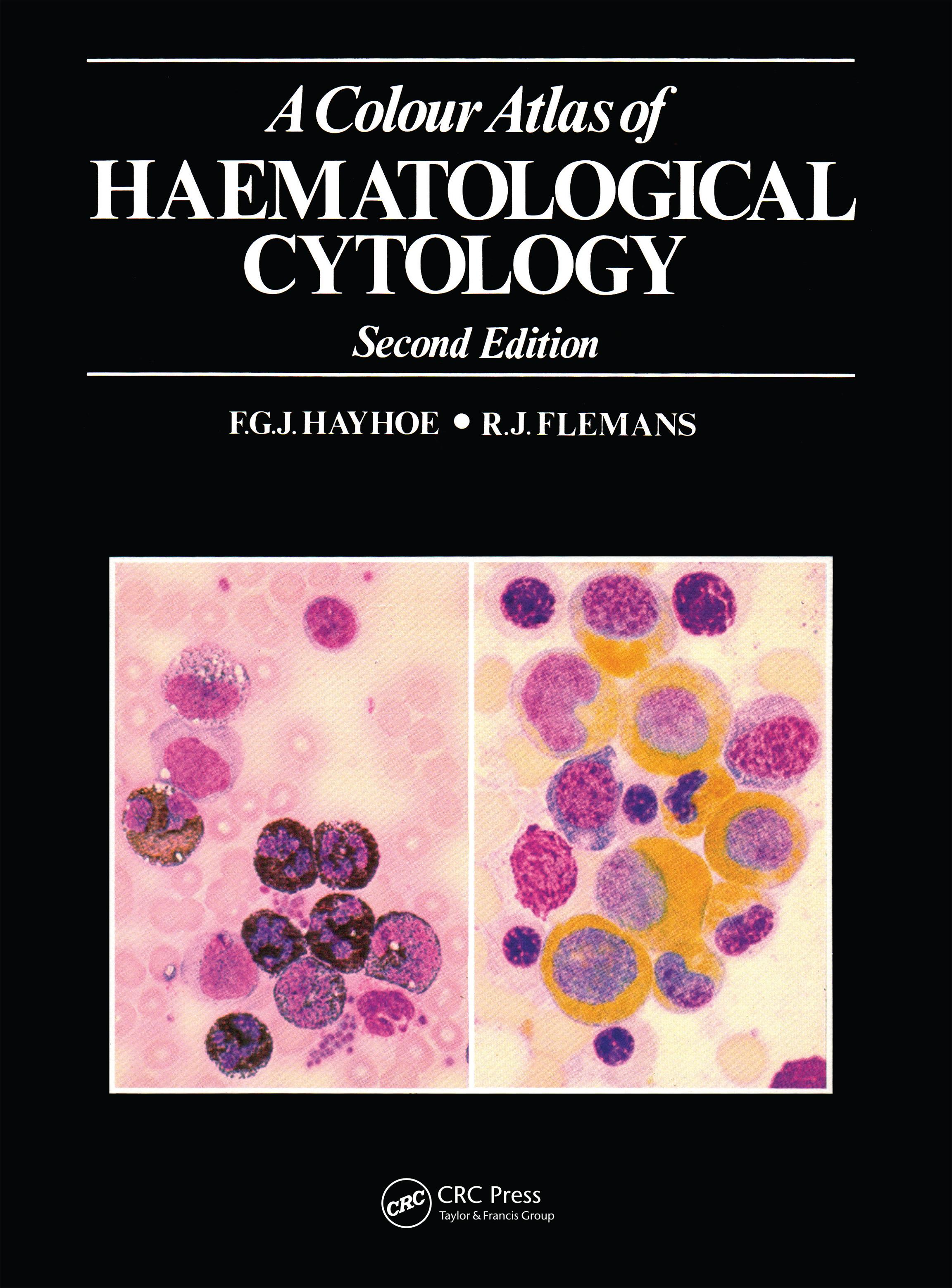
The second edition of this atlas has been revised and greatly en-larged to take account of recent diagnostic advances in the field of haematology. It contains 750 color photomicrographs of common and less common cell types, and is intended to supplement the per-sonal study of preparations under the microscope.
The photographs are grouped into five sections covering red cells and their precursors; nonerythroid cells of myeloid origin; lympho-cytes, plasma cells and their derivatives and precursors; miscella-neous cells of blood and marrow, including foreign cells and para-sites; and the imprint cytology of lymph nodes and spleen. Each section is preceded by a brief introduction, and an appendix pro-vides technical details of staining methods. The text accompanying the illustrations serves chiefly to identify the cells, but also gives explanations of certain unusual appearances and the cytochemical reactions where necessary.
A Colour Atlas of Haematological Cytology will be useful to students of medicine and medical laboratory science, as well as to doctors and technicians specializing in the diagnosis of haematological disease.
Betal nemt med kort, Klarna, Apple Pay eller Google Pay. Ikke tilfreds? Du har altid 14 dages fortrydelsesret. Læs mere i vores vilkår. Har du spørgsmål, så send os en mail på hello@memmo.org.
Memmo gør det nemmere at studere – uanset hvor du er i verden. Hos os samler du dine kursusbøger og smarte studieværktøjer ét sted: resuméer, quizzer, podcasts og flashcards. Og så er der Ted, din studieven, der svarer på alt, du undrer dig over. Over 50 000 studerende studerer allerede her – bygget til at hjælpe dig med at lære hurtigere og stresse mindre.
Du kan måske også lide
Køb bogen0